
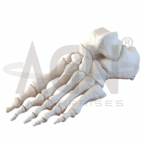
The Bones of the Foot Model from AGN Enterprises is a detailed anatomical teaching aid designed to demonstrate the skeletal anatomy and arrangement of the human foot. The model provides a three-dimensional representation of the tarsal, metatarsal, and phalangeal bones, helping students understand their positions, articulations, and contribution to foot structure and movement.
It is suitable for medical colleges, nursing institutes, physiotherapy colleges, orthopedic departments, podiatry programs, paramedical institutions, anatomy laboratories, and health-science training centers.
The Bones of the Foot Model demonstrates the major bones forming the human foot.
Depending on the specific model configuration, represented structures may include:
Talus
Calcaneus
Navicular
Cuboid
Medial Cuneiform
Intermediate Cuneiform
Lateral Cuneiform
Metatarsals
Phalanges
The exact mounting, articulation, flexibility, dimensions, and anatomical details may vary according to the supplied model.
The human foot normally contains 26 bones, organized into three major groups:
7 Tarsal Bones
5 Metatarsal Bones
14 Phalanges
Together, these bones form a strong yet mobile framework that supports body weight, absorbs forces, maintains balance, and contributes to walking and other movements.
The seven tarsal bones form the posterior portion of the foot.
They include:
The model helps students identify each tarsal bone and understand its relationship with adjacent structures.
The talus is positioned between the bones of the lower leg and the remaining foot skeleton.
It articulates with the tibia and fibula at the ankle and transfers forces from the leg into the foot.
The model provides a clear reference for understanding its central role in ankle and hindfoot anatomy.
The calcaneus is the largest tarsal bone and forms the structural foundation of the heel.
It supports substantial loads during standing, walking, and running.
The model helps learners examine its position beneath the talus and its relationship with other tarsal structures.
The navicular is located on the medial side of the midfoot, while the cuboid lies more laterally.
Both bones participate in important articulations connecting the hindfoot with the more distal regions of the foot.
Their positions can be clearly compared using the three-dimensional model.
The foot contains three cuneiform bones:
Medial Cuneiform
Intermediate Cuneiform
Lateral Cuneiform
These bones lie between the navicular and selected metatarsals and contribute to the structural organization and arches of the foot.
The foot contains five metatarsal bones, numbered I to V from the medial side toward the lateral side.
Each metatarsal consists generally of a base, shaft, and head.
The model helps students understand how the metatarsals connect the tarsal region with the toes.
The toes contain 14 phalanges.
The great toe, or hallux, normally contains two phalanges:
Proximal Phalanx
Distal Phalanx
The remaining four toes normally contain three each:
Proximal Phalanx
Middle Phalanx
Distal Phalanx
The model makes their arrangement easy to identify during practical anatomy lessons.
The arrangement of foot bones contributes to the formation of important anatomical arches, including the medial longitudinal, lateral longitudinal, and transverse arches.
These arches contribute to weight distribution, shock absorption, stability, and efficient movement.
The model provides a useful skeletal reference for explaining the structural basis of these arches.
The bones of the foot work together with joints, ligaments, muscles, and tendons to provide both stability and mobility.
They contribute to standing, balance, walking, running, propulsion, and adaptation to different surfaces.
Studying the bones in three dimensions helps students connect skeletal anatomy with foot biomechanics.
The Bones of the Foot Model provides students with a practical three-dimensional approach to studying foot anatomy.
Teachers can use it for lessons involving tarsal bones, metatarsals, phalanges, foot arches, ankle relationships, skeletal anatomy, and lower-limb biomechanics.
It supports bone identification, spatial visualization, practical demonstrations, and examination preparation.
AGN Enterprises supplies skeletal models, foot models, joint models, anatomical teaching aids, scientific instruments, and educational laboratory equipment for medical colleges, nursing institutes, physiotherapy programs, universities, and professional training centers.
Our anatomical teaching models provide clear three-dimensional representations for practical musculoskeletal and human anatomy education.









