
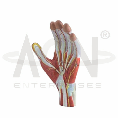
The Anatomy of the Hand, 3 Parts Model from AGN Enterprises is a detailed anatomical teaching aid designed to demonstrate the bones, muscles, tendons, ligaments, nerves, blood vessels, and other important structures of the human hand. Its three-part construction allows selected components to be separated for closer examination of superficial and deeper anatomy.
The model is suitable for medical colleges, nursing institutes, physiotherapy colleges, occupational therapy programs, orthopedic departments, anatomy laboratories, paramedical institutions, and health-science training centers.
The Anatomy of the Hand, 3 Parts Model provides a three-dimensional representation of important structures responsible for hand movement, stability, sensation, and function.
Depending on the specific configuration, represented structures may include:
Carpal Bones
Metacarpal Bones
Phalanges
Muscles
Tendons
Ligaments
Nerves
Blood Vessels
Palmar Structures
Dorsal Structures
The exact anatomical structures and removable components may vary according to the supplied model.
The three-part construction provides improved access to selected anatomical layers and structures.
Removable components allow students to examine represented structures individually and understand their relationships within the hand.
This design is particularly useful for practical demonstrations where deeper anatomical features need to be studied alongside superficial structures.
The adult human hand contains 27 bones, divided into three principal groups:
Carpals – 8 bones
Metacarpals – 5 bones
Phalanges – 14 bones
These bones provide the structural framework required for the wide range of movements performed by the hand and fingers.
The hand contains intrinsic muscles, while several tendons entering the hand originate from muscles in the forearm.
Together, these structures contribute to movements such as finger flexion, extension, abduction, adduction, and thumb opposition.
Where represented, the model helps students trace tendons and understand their relationships with surrounding bones and muscles.
Important nerves associated with hand function include branches of the median, ulnar, and radial nerves.
These nerves contribute to motor control and sensation in different regions of the hand. Blood is supplied through arterial networks primarily associated with branches of the radial and ulnar arteries.
Where represented, these structures provide useful references for clinical and functional anatomy.
Numerous joints and ligaments contribute to the mobility and stability of the hand.
The model can help students understand the relationships between the wrist, metacarpophalangeal joints, interphalangeal joints, bones, tendons, and supporting ligaments.
This makes it particularly useful for physiotherapy, orthopedics, and musculoskeletal education.
The Anatomy of the Hand, 3 Parts Model can support lessons on common hand movements and functional anatomy, including:
The model provides students with a three-dimensional approach to studying complex hand anatomy.
Teachers can use it for lessons involving hand bones, muscles, tendons, ligaments, nerves, blood vessels, joints, biomechanics, physiotherapy, and orthopedic anatomy.
Its three-part design supports anatomical identification, spatial visualization, practical demonstrations, and examination preparation.
AGN Enterprises supplies anatomical models, skeletal models, joint models, muscular teaching aids, scientific instruments, and educational laboratory equipment for medical colleges, nursing institutes, physiotherapy programs, paramedical institutions, and professional training centers.
Our anatomical models provide clear three-dimensional representations for practical and classroom-based human anatomy education.









