
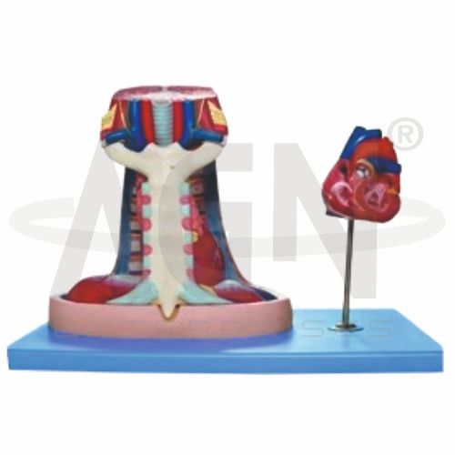
The Mediastinum Model from AGN Enterprises is a detailed anatomical teaching aid designed to demonstrate the central compartment of the thoracic cavity and the anatomical relationships of its major organs and structures. It helps students visualize the position of the heart, major blood vessels, trachea, bronchi, esophagus, and other mediastinal structures between the two lungs.
This educational model is suitable for medical colleges, nursing institutes, paramedical institutions, anatomy laboratories, respiratory therapy programs, and other health-science training centers.
The mediastinum is the central region of the thoracic cavity located between the right and left pleural cavities. It extends generally from the sternum anteriorly to the vertebral column posteriorly and from the superior thoracic aperture to the diaphragm.
Depending on the specific model configuration, structures represented may include:
Heart
Pericardium
Trachea
Main Bronchi
Esophagus
Aorta and Major Branches
Superior and Inferior Vena Cava
Pulmonary Vessels
Associated Nerves and Vessels
The exact structures and level of detail may vary according to the supplied model.
For anatomical study, the mediastinum is commonly divided into superior and inferior mediastinal regions.
The inferior mediastinum is further divided into:
Anterior Mediastinum
Middle Mediastinum
Posterior Mediastinum
This anatomical teaching aid can help students understand these divisions and relate them to the structures occupying each region.
The heart, enclosed by the pericardium, occupies much of the middle mediastinum.
The Mediastinum Model can support identification of important cardiovascular structures such as the aorta, venae cavae, pulmonary vessels, and other major vessels where represented.
This provides students with a useful three-dimensional perspective on the relationship between the heart and surrounding thoracic structures.
The trachea passes through the mediastinal region before dividing into the right and left main bronchi.
The model can help learners visualize the relationship between the tracheobronchial tree, heart, major vessels, and lungs.
This makes it useful for lessons involving thoracic anatomy, respiratory anatomy, and bronchial organization.
The esophagus travels through the thorax toward the diaphragm and lies posterior to several important mediastinal structures.
Where represented, students can use the model to examine its relationship with the trachea, heart, aorta, vertebral column, and surrounding structures.
This is particularly useful for understanding the spatial organization of the posterior thoracic region.
This anatomical teaching aid provides a three-dimensional approach to a region that can be difficult to understand from sectional diagrams alone.
Teachers can use it for lessons involving thoracic anatomy, cardiovascular anatomy, respiratory anatomy, mediastinal divisions, major blood vessels, tracheobronchial anatomy, and esophageal anatomy.
The model supports anatomical identification, spatial visualization, understanding of organ relationships, and practical classroom demonstrations.
AGN Enterprises supplies anatomical models, pathological models, biological teaching aids, and educational laboratory equipment for medical colleges, nursing institutes, paramedical institutions, schools, and professional training centers.
Our anatomical teaching models help learners understand complex human-body regions through clear visual and three-dimensional representations.









