
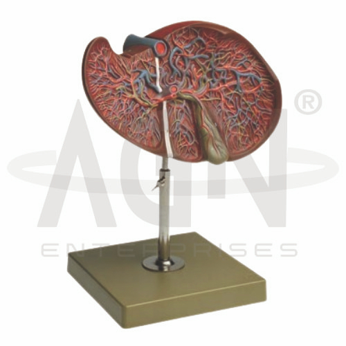
The Liver Section with Gallbladder Model from AGN Enterprises is a detailed anatomical teaching aid designed to demonstrate the internal structure of the human liver together with the gallbladder and associated biliary structures. The sectional presentation helps students understand liver anatomy and the anatomical relationship between the liver, gallbladder, bile ducts, and major vessels.
This educational model is suitable for medical colleges, nursing institutes, paramedical institutions, biology laboratories, anatomy departments, gastroenterology education, and health-science training centers.
The liver is a major abdominal organ involved in numerous metabolic and digestive functions. It processes nutrients, stores important substances, participates in detoxification, and produces bile.
Depending on the specific model configuration, structures represented may include:
Right Lobe
Left Lobe
Internal Liver Tissue
Gallbladder
Hepatic Ducts
Common Bile Duct
Hepatic Portal Vein
Hepatic Artery
Major Hepatic Vessels
The exact anatomical details may vary according to the supplied model.
A major advantage of this teaching model is its sectional representation of the liver.
Unlike a model showing only the external surface, the sectional design helps students visualize internal structures and understand how vessels and ducts are arranged within the organ.
This makes the model useful for studying both gross liver anatomy and hepatobiliary organization.
The Liver Section with Gallbladder Model clearly supports the study of the relationship between the liver and gallbladder.
The gallbladder is a small, pear-shaped organ located on the visceral surface of the liver. Its primary functions are to store and concentrate bile produced by the liver.
During digestion, bile is released into the biliary duct system and ultimately reaches the duodenum.
Where represented, students can use the model to trace the pathway of bile through the associated ducts.
A simplified pathway can be explained as:
Liver → Hepatic Ducts → Common Hepatic Duct → Common Bile Duct → Duodenum
The cystic duct connects the gallbladder with the biliary duct system, allowing bile to enter and leave the gallbladder.
This makes the model particularly useful for lessons involving bile production, storage, and transport.
The liver has a distinctive dual blood supply.
The hepatic artery supplies oxygenated blood, while the hepatic portal vein carries nutrient-rich blood from much of the digestive tract and associated organs to the liver.
Where these vessels are represented, the sectional model helps learners understand their relationship to the internal liver structures.
This anatomical teaching aid provides students with a three-dimensional approach to studying both the external and internal organization of the liver.
Teachers can use it for lessons involving liver anatomy, gallbladder anatomy, hepatobiliary system, bile ducts, digestive physiology, abdominal anatomy, and gastroenterology.
The sectional format supports anatomical identification, spatial visualization, understanding of internal structures, and practical classroom demonstrations.
AGN Enterprises supplies anatomical models, pathological models, biological teaching aids, and educational laboratory equipment for medical colleges, nursing institutes, paramedical institutions, schools, and professional training centers.
Our anatomical teaching models provide clear visual representations that help learners understand complex human organs and body systems.









