
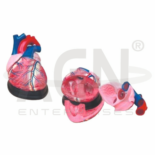
The Jumbo Heart Model from AGN Enterprises is an enlarged anatomical teaching aid designed to demonstrate the external and internal structures of the human heart in greater detail than a standard-size model. Its oversized construction makes important cardiac structures easier to identify during classroom demonstrations, practical anatomy sessions, and group teaching.
The model helps students study the atria, ventricles, heart valves, major blood vessels, septa, and coronary structures, depending on the supplied configuration. It is suitable for medical colleges, nursing institutes, paramedical institutions, schools, biology laboratories, anatomy departments, cardiology education, and health-science training centers.
The human heart is a muscular organ responsible for pumping blood through the pulmonary and systemic circulations.
Depending on the specific model configuration, structures represented may include:
Right Atrium
Left Atrium
Right Ventricle
Left Ventricle
Interventricular Septum
Tricuspid Valve
Mitral Valve
Pulmonary Valve
Aortic Valve
Aorta
Pulmonary Artery
Pulmonary Veins
Superior and Inferior Vena Cava
Coronary Blood Vessels
The exact anatomical details and removable components may vary according to the supplied model.
A major advantage of the Jumbo Heart Model is its enlarged construction. Structures that can be difficult to observe on smaller models become easier to demonstrate to groups of students.
The oversized representation is particularly useful for explaining the arrangement of the chambers, valves, major vessels, and internal cardiac structures.
It can also help learners develop a clearer understanding of the orientation of the heart within the cardiovascular system.
The heart consists of four chambers:
Right Atrium – receives deoxygenated blood returning from the body.
Right Ventricle – pumps blood toward the lungs.
Left Atrium – receives oxygenated blood returning from the lungs.
Left Ventricle – pumps oxygenated blood into the systemic circulation.
The enlarged model helps students visualize the relative positions and wall structures of these chambers.
The valves of the heart help maintain one-way blood flow.
The four principal valves are:
Tricuspid Valve
Pulmonary Valve
Mitral (Bicuspid) Valve
Aortic Valve
Where internal sections are accessible, learners can examine the position of these valves in relation to the atria, ventricles, and major blood vessels.
The model can help students identify the major vessels connected to the heart.
These include the aorta, pulmonary trunk and arteries, pulmonary veins, superior vena cava, and inferior vena cava.
Studying these structures together helps learners understand how the heart connects pulmonary and systemic circulation.
Teachers can use this anatomical teaching aid to demonstrate the basic pathway of blood:
Body → Venae Cavae → Right Atrium → Right Ventricle → Pulmonary Arteries → Lungs → Pulmonary Veins → Left Atrium → Left Ventricle → Aorta → Body
The enlarged format makes this pathway particularly suitable for classroom demonstrations and introductory cardiovascular education.
Where represented, the model may also demonstrate the major coronary blood vessels located on the surface of the heart.
These vessels supply the heart muscle itself with oxygenated blood and nutrients. Their inclusion supports lessons involving cardiac anatomy, coronary circulation, and introductory clinical cardiology.
This enlarged anatomical teaching aid helps students understand complex cardiac anatomy through direct three-dimensional observation.
It is useful for teaching heart anatomy, cardiovascular physiology, cardiac chambers, valves, blood circulation, coronary vessels, and cardiopulmonary relationships.
The jumbo format is especially valuable for group demonstrations because anatomical structures can be viewed more clearly than on smaller heart models.
AGN Enterprises supplies anatomical models, pathological models, biological teaching aids, and educational laboratory equipment for schools, medical colleges, nursing institutes, paramedical institutions, and professional training centers.
Our enlarged anatomical teaching models help make complex human-body structures easier to visualize during practical and classroom education.









