
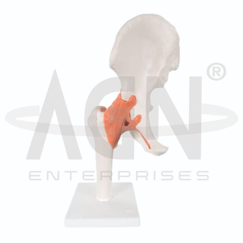
The Hip Joint Model from AGN Enterprises is a detailed anatomical teaching aid designed to demonstrate the bones, ligaments, and structural relationships of the human hip joint. The three-dimensional model helps students understand how the head of the femur articulates with the acetabulum of the pelvis to form a strong and mobile weight-bearing joint.
It is suitable for medical colleges, nursing institutes, physiotherapy colleges, orthopedic departments, paramedical institutions, anatomy laboratories, sports-science programs, and health-science training centers.
The Hip Joint Model provides a clear representation of the major anatomical structures involved in the hip region.
Depending on the specific model configuration, represented structures may include:
Head of Femur
Neck of Femur
Acetabulum
Pelvic Bone
Joint Capsule
Iliofemoral Ligament
Pubofemoral Ligament
Ischiofemoral Ligament
Associated Joint Structures
The exact anatomical structures, movable components, and level of detail may vary according to the supplied model.
The hip is a ball-and-socket synovial joint formed between the rounded head of the femur and the acetabulum of the pelvis.
The deep acetabulum provides substantial stability while allowing movement in several directions.
The model helps students visualize the relationship between the proximal femur and pelvic bones more clearly than flat anatomical illustrations.
The rounded femoral head fits within the acetabulum to form the principal articulation of the hip.
The femoral neck connects the head with the shaft of the femur and is an important region in clinical and orthopedic anatomy.
The model provides a useful three-dimensional reference for studying the orientation of these structures.
Strong ligaments reinforce the hip joint and contribute to its stability.
Important ligaments include:
Iliofemoral Ligament
Pubofemoral Ligament
Ischiofemoral Ligament
Where represented, the model helps learners identify these ligaments and understand how they support the joint during standing and movement.
The Hip Joint Model can be used to demonstrate or explain the major movements possible at the hip, including:
The range of movement available on the physical model depends on its specific construction.
The hip combines mobility with substantial structural stability.
The deep acetabulum, surrounding joint capsule, strong ligaments, and associated muscles contribute to maintaining the femoral head within the socket.
Understanding these relationships is particularly important for physiotherapy, orthopedics, sports medicine, and biomechanics education.
The model can support explanations of clinically important topics involving the hip region, such as femoral-neck fractures, joint degeneration, dislocation, ligament function, and joint replacement concepts.
It is intended as an educational anatomical reference rather than a diagnostic tool.
The Hip Joint Model provides students with a three-dimensional approach to studying joint anatomy and biomechanics.
Teachers can use it for lessons involving hip anatomy, pelvic anatomy, femur, ligaments, joint movement, musculoskeletal anatomy, physiotherapy, biomechanics, and orthopedics.
The model supports anatomical identification, spatial visualization, practical demonstrations, and examination preparation.
AGN Enterprises supplies anatomical models, skeletal models, joint models, muscular teaching aids, scientific instruments, and educational laboratory equipment for medical colleges, nursing institutes, physiotherapy programs, paramedical institutions, and professional training centers.
Our anatomical teaching aids provide clear three-dimensional representations for practical and classroom-based human anatomy education.









