
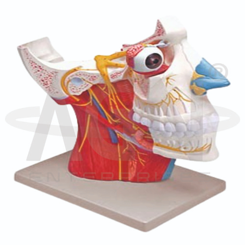
The Cranial and Autonomic Nerves Model from AGN Enterprises is a detailed anatomical teaching aid designed to demonstrate the cranial nerves and important components of the autonomic nervous system. The model helps students understand nerve distribution, anatomical pathways, and relationships between the brain, head, neck, and autonomic structures.
It is suitable for medical colleges, nursing institutes, physiotherapy colleges, neuroscience departments, paramedical institutions, anatomy laboratories, and health-science training centers.
The Cranial and Autonomic Nerves Model provides a three-dimensional representation of selected structures of the peripheral and autonomic nervous systems.
Depending on the specific model configuration, represented structures may include:
Cranial Nerves
Brain and Brainstem
Sympathetic Nerves
Parasympathetic Nerves
Autonomic Ganglia
Cervical Nerve Structures
Head and Neck Nerve Pathways
Associated Neural Structures
The exact nerves, pathways, colors, removable components, and anatomical details may vary according to the supplied model.
The human nervous system contains 12 pairs of cranial nerves, conventionally numbered I to XII:
I – Olfactory Nerve
II – Optic Nerve
III – Oculomotor Nerve
IV – Trochlear Nerve
V – Trigeminal Nerve
VI – Abducens Nerve
VII – Facial Nerve
VIII – Vestibulocochlear Nerve
IX – Glossopharyngeal Nerve
X – Vagus Nerve
XI – Accessory Nerve
XII – Hypoglossal Nerve
Where represented, the model helps students identify these nerves and understand their anatomical relationships.
The olfactory nerve (CN I) is associated with the sense of smell, while the optic nerve (CN II) carries visual information from the retina toward the brain.
These nerves provide important examples of cranial nerves with specialized sensory functions.
Where represented, students can trace their relationship with the brain and sensory structures.
Cranial nerves III, IV, and VI are closely associated with movements of the eyes.
The oculomotor, trochlear, and abducens nerves innervate specific extraocular muscles and contribute to coordinated eye movement.
The model can support demonstrations of their pathways and functional relationships.
The trigeminal nerve (CN V) is a major sensory nerve of the face and also provides motor innervation to muscles involved in mastication.
The facial nerve (CN VII) supplies muscles of facial expression and carries additional sensory and autonomic fibers.
Where represented, the model helps learners compare their distribution in the facial region.
The vagus nerve (CN X) has an especially important relationship with the autonomic nervous system.
It provides extensive parasympathetic innervation to structures in the thorax and abdomen and also has sensory and motor functions.
The model can help students understand why the vagus nerve is important in both cranial-nerve and autonomic-system studies.
The autonomic nervous system (ANS) regulates many involuntary physiological functions.
Its major divisions include:
Sympathetic Division
Parasympathetic Division
These systems contribute to regulation of functions involving the heart, blood vessels, digestive tract, glands, pupils, and other internal structures.
The sympathetic division helps coordinate physiological responses that prepare the body for increased activity.
Where represented, the model may demonstrate sympathetic trunks, ganglia, and associated nerve pathways.
This allows students to connect anatomical structures with autonomic nervous-system functions.
The parasympathetic division contributes to functions associated with rest, digestion, energy conservation, and regulation of internal organs.
Several cranial nerves carry parasympathetic fibers, particularly cranial nerves III, VII, IX, and X.
The model provides a useful visual reference for studying these functional relationships.
The Cranial and Autonomic Nerves Model provides students with a three-dimensional approach to studying complex neural pathways.
Teachers can use it for lessons involving cranial nerves, sympathetic nerves, parasympathetic nerves, autonomic ganglia, brainstem anatomy, nerve pathways, and neuroanatomy.
It supports nerve identification, spatial visualization, practical demonstrations, and examination preparation.
AGN Enterprises supplies neurological models, nervous-system models, brain models, anatomical teaching aids, scientific instruments, and educational laboratory equipment for medical colleges, nursing institutes, physiotherapy programs, universities, and professional training centers.
Our anatomical models provide clear three-dimensional representations for practical neuroanatomy and medical education.









