
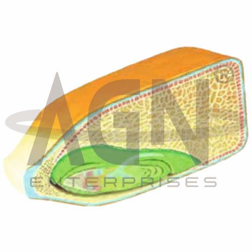
The Corn Seed Model, 2 Parts from AGN Enterprises is an enlarged botanical teaching model designed to demonstrate the internal and external anatomy of a maize (corn) seed. Its two-part construction allows the model to be opened for clear observation of the embryo, endosperm, seed covering, and other important structures.
The model is suitable for schools, colleges, universities, botany laboratories, biology departments, agricultural institutes, horticulture programs, and plant-science training centers.
The Corn Seed Model, 2 Parts provides an enlarged three-dimensional representation of a corn grain, botanically known as a caryopsis.
Depending on the specific model configuration, represented structures may include:
Pericarp and Seed Coat
Endosperm
Embryo
Scutellum
Plumule
Radicle
Coleoptile
Coleorhiza
Embryonic Axis
The exact structures, colors, magnification, and sectional details may vary according to the supplied model.
The model is designed in two separable parts, allowing students to open the corn seed and examine its internal organization.
This sectional design provides a clearer understanding of structures that cannot normally be observed easily from the exterior of an intact seed.
The two-part format is especially useful for classroom demonstrations and practical botany lessons.
Corn or maize (Zea mays) is a monocotyledonous flowering plant.
Its grain is technically a caryopsis, a dry fruit in which the fruit wall is closely associated with the seed coat.
The model helps students understand the characteristic organization of a maize grain and distinguish its major internal regions.
The endosperm forms a large portion of the mature corn grain.
It contains stored nutrients that support the developing embryo and early seedling during germination.
The enlarged model clearly demonstrates the relative size and position of the endosperm compared with the embryo.
The embryo represents the young developing plant within the seed.
It contains structures that eventually develop into the shoot and root systems during germination.
The model provides a three-dimensional reference for locating the embryo relative to the endosperm.
The scutellum is the single cotyledon characteristic of the grass embryo.
It lies between the embryonic axis and the endosperm and participates in the transfer of stored nutrients to the developing embryo during germination.
Its presence is an important feature when studying monocot seed anatomy.
The plumule is the embryonic shoot that develops into the above-ground portion of the young plant.
In maize, it is protected by a specialized sheath called the coleoptile.
Where represented, the model helps students distinguish the developing shoot from surrounding embryonic structures.
The radicle is the embryonic root and contributes to the initial root development during germination.
In maize, the radicle is enclosed by a protective structure known as the coleorhiza.
The model helps learners understand the positions of these structures within the embryo.
The Corn Seed Model, 2 Parts can be used to demonstrate important features associated with monocot seed anatomy, including:
These characteristics make the model useful for comparisons with dicot seeds.
The model can support lessons on maize seed germination by helping students identify structures involved in the development of the young plant.
During germination, stored nutrients support embryonic growth while the radicle and shoot structures begin developing into the root and aerial systems.
The physical model helps connect seed anatomy with the germination process.
The Corn Seed Model, 2 Parts provides an enlarged three-dimensional approach to studying seed anatomy.
Teachers can use it for lessons involving maize seed structure, monocot seeds, embryo anatomy, endosperm, scutellum, plumule, radicle, coleoptile, coleorhiza, and seed germination.
It supports structural identification, practical demonstrations, comparative plant anatomy, and examination preparation.
AGN Enterprises supplies botanical models, plant-anatomy models, biological teaching aids, scientific instruments, and educational laboratory equipment for schools, colleges, universities, agricultural institutes, and professional laboratories.
Our botanical teaching models provide clear three-dimensional representations to support practical botany and biology education.









