
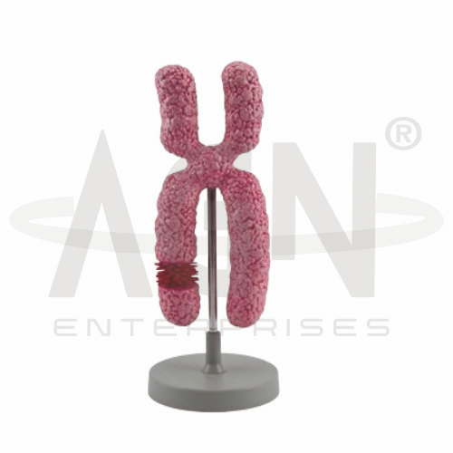
The Chromosome Model from AGN Enterprises is a three-dimensional biological teaching aid designed to demonstrate the structure and organization of a chromosome. Its enlarged representation makes chromosome morphology easier to visualize and helps students understand important concepts related to chromatids, centromeres, DNA organization, genetics, and cell division.
The model is suitable for schools, colleges, universities, medical colleges, nursing institutes, biology laboratories, genetics laboratories, cytology departments, and health-science training centers.
Chromosomes are organized structures composed primarily of DNA associated with proteins. They carry genetic information required for cellular function, growth, development, and reproduction.
Depending on the specific model configuration, represented features may include:
Sister Chromatids
Centromere
Chromosome Arms
Chromosomal Ends
Constriction Region
Duplicated Chromosome Form
The exact structural details, scale, and representation may vary according to the supplied model.
A duplicated chromosome consists of two sister chromatids produced during DNA replication.
The chromatids contain corresponding copies of genetic information and remain associated until they separate during the appropriate stage of cell division.
The enlarged model helps students visualize the relationship between the two chromatids.
The centromere is the constricted chromosomal region where sister chromatids remain closely associated.
During cell division, protein structures assembled around the centromeric region participate in chromosome movement and segregation.
The model provides a clear visual reference for identifying the centromere and understanding its position within a chromosome.
The centromere divides each chromatid into chromosome arms.
These are commonly described as:
p Arm – Short Arm
q Arm – Long Arm
The relative lengths of these arms depend on the position of the centromere and contribute to chromosome classification.
Chromosomes provide an organized method for packaging long DNA molecules within cells.
DNA associates with proteins to form chromatin, which undergoes increasing condensation as cells prepare for division. Highly condensed chromosomes become readily distinguishable during particular stages of cell division.
The model helps connect DNA organization with visible chromosome morphology.
The Chromosome Model can be used to explain chromosome behavior during mitosis and meiosis.
During cell division, duplicated chromosomes undergo organized movement and segregation so that genetic material can be distributed to resulting cells.
This makes the model useful for lessons involving genetics, reproduction, heredity, and cellular biology.
Chromosomes contain genes that contribute to inherited biological characteristics.
Humans normally have 46 chromosomes arranged in 23 pairs in most somatic cells. One chromosome of each pair is inherited from each biological parent.
The model provides a useful starting point for explaining chromosomes, genes, DNA, and heredity.
The Chromosome Model provides students with a three-dimensional alternative to studying chromosome structure solely through textbook diagrams.
Teachers can use it for lessons involving chromosome anatomy, chromatids, centromeres, DNA, genes, mitosis, meiosis, genetics, heredity, and cell biology.
The enlarged representation supports visual learning, practical demonstrations, concept development, and examination preparation.
AGN Enterprises supplies biological models, anatomical teaching aids, scientific instruments, laboratory equipment, and educational products for schools, colleges, universities, medical institutions, and professional training centers.
Our biological teaching models provide clear three-dimensional representations to support practical science and laboratory education.









