
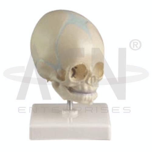
The Baby Skull Model from AGN Enterprises is a detailed anatomical teaching aid designed to demonstrate the characteristic anatomy of an infant skull, including the cranial bones, sutures, fontanelles, facial skeleton, and developing cranial structure.
The model is particularly useful for comparing the infant skull with the adult skull and understanding the anatomical adaptations that permit brain growth and changes in skull shape during early development.
It is suitable for medical colleges, nursing institutes, pediatric departments, midwifery programs, anatomy laboratories, paramedical institutions, biology departments, and health-science training centers.
The Baby Skull Model provides a three-dimensional representation of important structures of the infant cranium and face.
Depending on the specific model configuration, represented structures may include:
Frontal Bone
Parietal Bones
Temporal Bones
Occipital Bone
Facial Bones
Cranial Sutures
Anterior Fontanelle
Posterior Fontanelle
Sphenoidal Fontanelles
Mastoid Fontanelles
Orbits
Nasal Cavity
Mandible
The exact structures, dimensions, movable components, colors, and anatomical details may vary according to the supplied model.
The infant skull differs considerably from the adult skull.
At birth and during early development, several cranial bones have not yet completely united. Flexible sutures and fontanelles occur between specific bones and accommodate changes associated with childbirth and rapid brain growth.
The model helps students visualize these developmental characteristics in three dimensions.
Fontanelles are membrane-covered spaces located between incompletely fused cranial bones in the developing infant skull.
Major fontanelles include:
These structures gradually close as skull development progresses.
The anterior fontanelle is the largest and most clinically recognizable fontanelle.
It is located near the junction of the frontal and parietal bones.
The model provides a useful visual reference for understanding its position, shape, and relationship with surrounding cranial bones.
The posterior fontanelle is located near the junction of the parietal bones and occipital bone.
It is considerably smaller than the anterior fontanelle and normally closes earlier during infant development.
Where clearly represented, students can compare the anterior and posterior fontanelles directly.
The infant skull contains important fibrous joints known as cranial sutures.
Major sutures represented may include the:
Coronal Suture
Sagittal Suture
Lambdoid Suture
Other developing sutural relationships may also be represented depending on the model.
The cranial bones surround and protect the developing brain.
The model helps students identify the frontal, parietal, temporal, and occipital regions and understand how these bones relate to the major sutures and fontanelles.
This is particularly useful when comparing infant and adult skull anatomy.
The facial portion of an infant skull has different proportions compared with an adult skull.
The cranial region is proportionally larger, while the facial skeleton is relatively smaller and continues developing throughout childhood.
The model provides a useful reference for studying these developmental differences.
The infant skull undergoes substantial growth as the brain and facial structures develop.
Sutures and fontanelles provide flexibility during early development and permit expansion of the cranial vault.
Over time, fontanelles close and the skull gradually develops more adult-like proportions.
The Baby Skull Model is especially useful for pediatric, obstetric, and midwifery anatomy education.
It can support lessons involving fontanelles, cranial sutures, infant head anatomy, skull development, childbirth-related anatomy, and differences between infant and adult skulls.
The model is intended for anatomical education rather than clinical diagnosis.
The Baby Skull Model provides students with a three-dimensional approach to studying infant cranial anatomy.
Teachers can use it for lessons involving infant skull bones, fontanelles, sutures, cranial development, facial anatomy, pediatric anatomy, and developmental anatomy.
It supports anatomical identification, comparative study, practical demonstrations, and examination preparation.
AGN Enterprises supplies skull models, skeletal models, pediatric anatomical models, human anatomy teaching aids, scientific instruments, and educational laboratory equipment for medical colleges, nursing institutes, universities, and professional training centers.
Our anatomical teaching models provide clear three-dimensional representations for practical and classroom-based human anatomy education.









